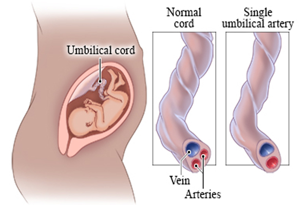
Single umbilical artery
The umbilical cord forms between 13 and 38 days after conception and normally serves as the conduit for two umbilical arteries and one umbilical vein. The normal umbilical cord contains 2 arteries and 1 vein (there vessel cord ) single umbilical artery is characterized by the absence of either the left- or right umbilical artery. This malformation has a reported incidence of 0.5-6% in singleton pregnancies it increases of 3-4 times in twin pregnancies
Single umbilical artery is of 3 types
TYPE 1– The entire length of the cord from baby to placenta , has just 2 vessels – one artery and one vein
TYPE 2– There are 3 vessel at the baby’s end of the cord but at the placental side, 2cms from the surface of the placenta, there are just 2 vessels .
The cause is anagtomosis.
TYPE 3– The cord initially has 3 vessels but occlusion close one artery of the cord .
It is related to circulatory problems in the umbilical cord and baby.
Sometimes there can be persistence of the original single allantoic artery of the baby stalk.
Neonates with SUA are at a higher risk of congenital anomalies and chromosomal abnormalities. The most common congenital anomalies associated with SUA are renal, hydrocephalus, thanatophoric dysplasia, gastroschisis, sacral agenesis hypospadias followed by cardiovascular and musculoskeletal.
There are three theories to explain how a SUA may form during development.
The first is that a primary agenesis of one umbilical artery results in a SUA. Another theory attributes the phenomenon to a secondary atrophy or atresia of a previously normal umbilical artery.
A third theory describes a persistence of the original allantoic artery of the body stalk as an explanation for SUA.
Embryological considerations, as well as the detection of occluded remnants of a second umbilical artery in some SUA fetuses, suggest that the second theory is the most likely explanation
Diagnosis
It can be detected in the first trimester of pregnancy with the use of 2D ultrasound. The sonographer is able to identify a 2 vessel cord in an image with the bladder and color Doppler, which will show only one artery going around one side of the bladder. In a normal fetus, there would be 2 arteries (one on each side of the bladder)
Management
When a 2 vessel cord is detected, a through search for other anomalies is required. A fetal 2D-ECHO is warranted.
Invasive testing is not recommended in isolated single umbilical artery.
Invasive testing with chromoromal evalvation (Micro array) is recommended if associated malformations are detected.
Serial growth scans with doppler indices are needed once a 2 vessel cord is seen
For normal weight fetures with isolated single umbilical artery and normal insertion of the cord, no particular precautions during labour are needed and induction can be performed
Studies have shown a higher incidence of cesarean section in these women ( due to prematurity, fetal growth restriction and oligohydramnios) .


